Explanation of Treatment Plan
Treatment protocol is based on the physiological facts how the body heals based on the three phases of healing after an injury:
Inflammatory Phase (time frame first 72hrs. after injury)
- During this phase, damaged cells are cleared out, along with bacteria and other pathogens or debris. This happens by phagocytosis where white blood cell’s eat debris by engulfing it. Platelet derived growth factors are released into the wound that cause migration and division of cells during the proliferative phase.
Repair Phase (time frame is 72 hours after injury up to 12 weeks)
- During this phase, angiogenesis, collagen deposition, granulation tissue formation, ephithelialization, and wound contraction occur. In angiogenesis, vascular endothelial cells form new blood vessels. In fibroplasia and granulation tissue formation, fibroblasts grow and form a new, provisional extracellular matrix by excreting collagen and fibronectin. Concurrently, re-epithelialization of the epidermis occurs, in which epithelial cells proliferate and “crawl” atop the wound bed, providing cover for the new tissue. In wound contraction, myofibroblasts decrease the size of the wound by gripping the wound edges and contracting using a mechanism that resembles that is smooth muscle cells. When the cells’ roles are close to complete, unneeded cells undergo apoptosis or cell death.
Remodel Phase (time frame is after 12 weeks up to 12 months)
- During maturation and remodeling, collagen is realigned along tension lines, and cells that are no longer needed are removed by programmed cell death, or apoptosis.
Time factors can vary on additional factors such as age, general health condition, use of medications and work and lifestyle activities that may cause re-injuries and may prolong the healing times. The existence and improvement of positive biomarkers will dictate the frequency and duration of care. An injury will naturally progress through the three phases of healing if there is no further irritant. Further irritation can initiate the inflammatory process with tissue that has progressed into the repair phase creating the inflammatory cycle while the repair cycle is in progress and tissue that has progressed into the remodel phase (separate and distinct cell damage). Any injury can have all three phases of healing present in cellular tissue if there is continual irritation or exacerbation. Depending on the irritant or exacerbation you can have all three phases of healing taking place simultaneously while previously injured tissue is in a separate and distinct phase at different intervals of time.
Treatment goals for the inflammatory phase are as follows:
- Decrease pain (intensity and frequency)
- Decrease inflammation (acute and chronic)
Treatment goals for the repair phase of healing are as follows:
- Repair type 3 collagen (scar tissue)
- Reduce muscle spasm
- Reduce nerve inflammation
- Correct abnormal joint biomechanics
- Increase flexibility and range of motion
- Improve functional restrictions (ADL’s)
The body produces type 3 collagen (scar tissue) in the injured area. The scar tissue lays down randomly and has 4 negative effects compared to healthy type 1 collagen:
- Less strength (up to 30% weaker)
- Less flexible (up to 30% less elastic)
- Increased number of peripheral nerve endings (get entangled in randomly laid scar tissue)
- Increased sensitivity of nerve endings (fire pain at lower threshold)
Illustration #5 shows the comparison of healthy collagen vs injured type 3 collagen (scar tissue).
ILLUSTRATION #5, TYPE 1 COLLAGEN AND TYPE 3 COLLAGEN
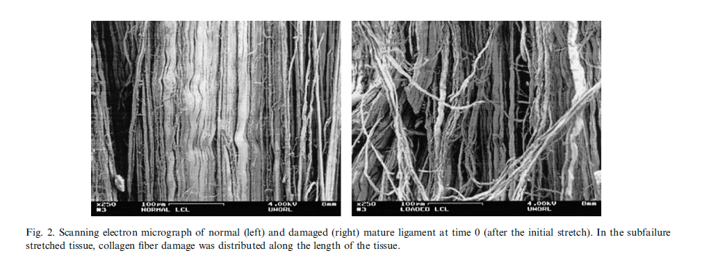
Without physical medicine (chiropractic, non-linear intersegmental and/or manual traction) the collagen lays down randomly and suffers these 4 negative characteristics. It is the stress at the 3rd range of motion (end joint play beyond gross active and passive ROM) that helps the collagen re-align.
Without physical medicine the patient would heal wrong and the likelihood of permanency is significantly increased making the patient more susceptible to re-injury under ordinary activities of daily living. The patient could also expect a decrease in the injured areas elasticity, resulting in less mobility or motion during activities of daily living. The patient would also experience the negative effects of peripheral nerve healing. During nerve re-genesis the body produces excessive nerve tissue that often becomes entangled in the new type 3 collagen which form randomly in crossing patterns. When nerve endings become entangled they are more prone to fire or cause pain during normal movement patterns. Also the nerves will fire at a lower threshold which increases the sensitivity of the nerve mean new nerve endings fire at a lower stimulus causing pain. If left untreated, the patient will end up with a permanent injury which includes pain with normal activities of daily living where none existed prior to the injury.
Treatment goals for the remodel phase of healing are as follows:
- Return to pre-injury status
- Reach MMI (maximum medical improvement)
- Implement active care to increase strength and elasticity
Therapeutic Protocol:
- Icing instructions to decrease inflammation
- Physical Medicine (Physiotherapy and exercise protocol) to create normal joint biomechanics
- Non-linear intersegmental and manual traction to assist in repairing type 3 collagen (scar tissue)
- Active care recommendations (exercises/stretches) to increase strength and elasticity
All forms of therapy can be used during the three phases of healing if the subjective and objective biomarkers are present and documented (i.e. – pain relief therapy during the remodel phase, active motion therapy for the repair phase during the remodel time frame).
Range of Motion with Overpressure is to test the spinal ligaments in the 3 ranges of motion of the spine, active/passive and movement into the para-physiological joint space. It is noted when pain is elicited in active/passive or end joint play. The following system is used rate the tolerance of the spinal ligaments in flexion, extension, left and right lateral flexion and left and right rotation:
3 – can’t perform active ROM without pain
2 – can’t get through full active and into passive ROM without pain
1 – can get through active and passive ROM but movement into the para-physiological joint
space causes pain (under load)
0 – can move the joint under load with no pain
Patient will be re-evaluated on a regular basis (4 weeks or 8-12 visits) to determine response to care with the following tools of assessment:
- Pain reassessments to determine level of decreased pain
- Post static x-rays to measure level of correction of abnormal joint biomechanics
- Functional Self Movement Screens – to determine quality movement and coordination of muscles during movements of ADL. (decrease chance of re-injury, falls ect.)
- Physical Capacity Test – to determine the strength (capacity) and ability of muscles to support body weight during ADL
- Outcome Assessment Tools Score (OATS) to determine improvement with functional capacities and ADL’s
- Active ROM testing to compare gross ROM with AMA norms and ROM with overpressure to test the intersegmental joint space under stress.
- Orthopedic tests to measure sensitivity to injured area including ROM with overpressure
Need for care is based on the following criteria in assessing the 3 degrees of an injury:
Subthreshold
- Quantity or quality of injured cells does NOT affect signs, symptoms or functional capacity. No POSTIVE objective biomarkers. This would not substantiate a need for care.
Functional Capacity
- Quantity or quality of injured cells DOES affect signs, symptoms of functional capacity such as ROM and sensitivity to pain. Subjective and objective biomarkers are present, requires treatment based on the three phases of healing and the sensitivity of the tests is important to objectify and quantify the injury.
Anatomical Failure
- Similar to functional capacity but at MMI will have higher likelihood of a degree of impairment not likely to improve with treatment.
Each re-exam it will be assessed if there are any POSITIVE biomarkers that substantiate the need for further care. Static MMI is determined based on where the patient is in regards to the 3 phases of healing and if there are two successive re-exams that show no indicators that further care will make a clinically significant change in the biomarkers. The cells will still go through the 3 phases of healing if the injury is documented, however the need for care for that injury during the 3 phases is based on the presence of positive biomarkers that substantiate the need for care through the 3 phases. The re-exams determine the trend-lines of response to care, overall treatment protocol is never based on an individual SOAP note or one re-exam on its own, and it is typical of injuries to have micro-trends of up and down days. Stability and MMI is determined based on a macro-trend of 2 successive re-exams in which stability is noted and how close the patient is back to normal or MMI has been reached.